About This Page
- Pictures with a © symbol were taken by me
- Click on images to view full size in lightbox
- This documents my accident and recovery from June 2005 to September 2006
June 2005: The Accident
The Last Resort, Aravis area of the Haute Savoie, French Alps
Annecy Hospital - 26th June, 2005 (Day 0):
- Fractured right knee
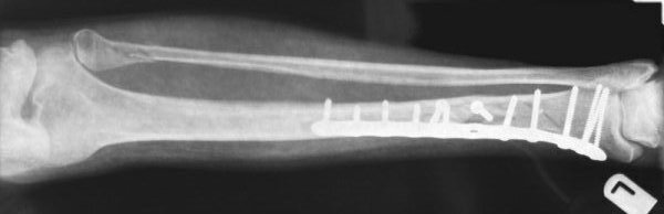
- Compound (small 1.5cm puncture) fracture to right tibia, and fractured right fibula
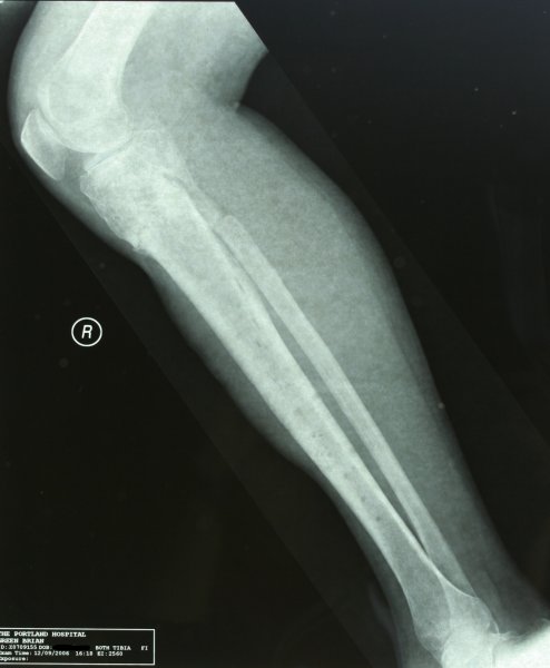
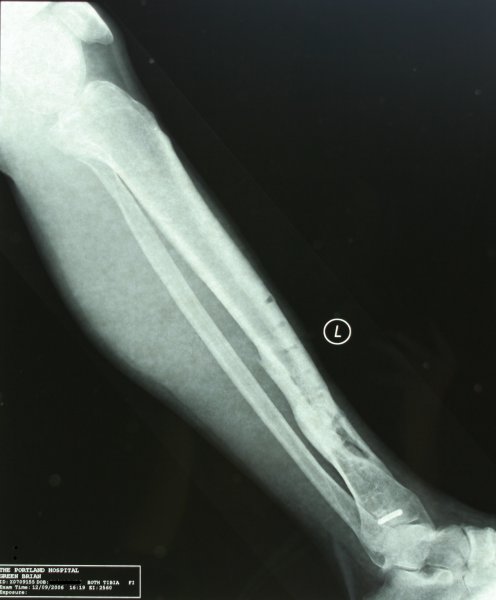
- Fracture to lower end of the left tibia, and fractured left fibula
Operation - 27th June, 2005 (Day 1):
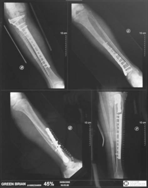
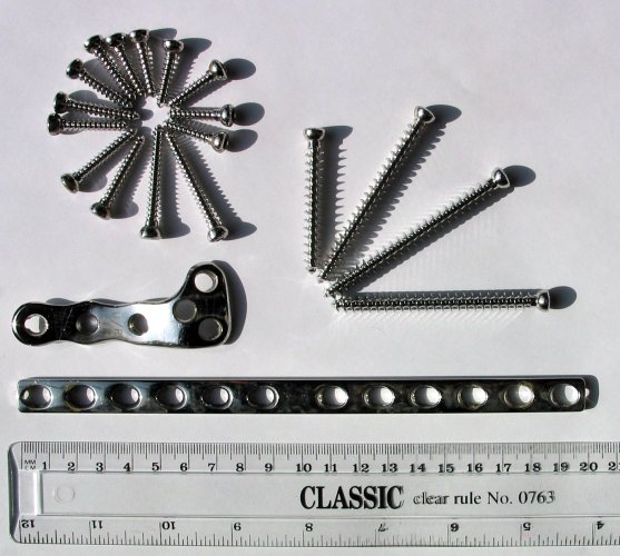
- Dr. Allamel: left tibia plate with 13 screws - 18cm long, 28 stitches
- Dr. Martinez: tibia right knee plate with 6 screws, right tibia plate with 12 screws - 39cm long, 43 staples and 1 stitch!
I'm flown back to the UK on 6th July (Day 10) by air ambulance (arranged with Specialty Group Ltd) directly to The London Clinic, to be monitored by Mr. M. Bankes (Consultant Orthopedic Surgeon).
Hydro/Physiotherapy, five days a week, started on the 11th July, 2005 (Day 15). Finally home on 3rd September, 2005 (Day 69) after 59 days at the clinic.

X-rays from Annecy
Physiotherapy Journey
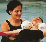
For the following 6 months, 2-3 times a week, I have physiotherapy at The London Clinic.
I'm extremely fortunate to have the multi-talented Janine, of the physiotherapy department at The London Clinic, as my physiotherapist throughout this period. I slowly re-learn how to walk, and balance on one foot on a rocker board whilst playing catch...
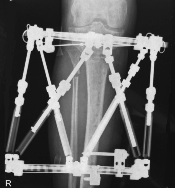
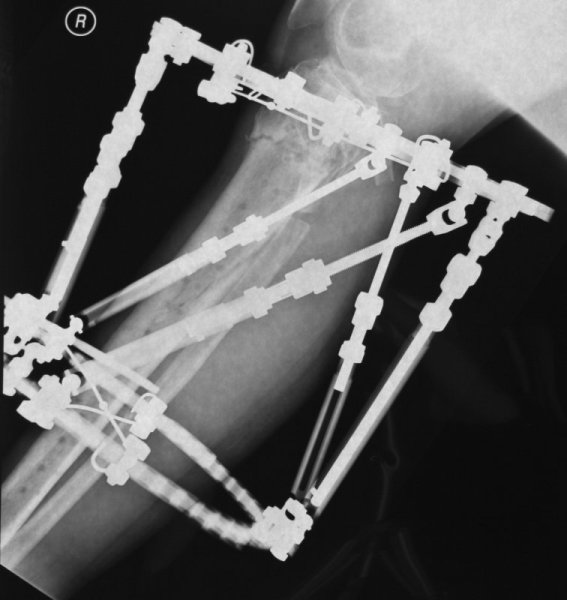
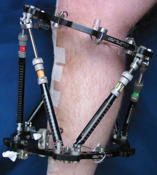
March 2006: Second Operation - Taylor Spatial Frame
Despite becoming almost fully mobile, and only requiring a stick for long journeys, there's still more to be done...
On 2nd March, 2006 (Day 249), at the Hospital of St. John & St Elizabeth (HJE), my right leg is operated on for the second time. The operation, 3.5 hours long, is done by Mr Hill, a UK surgeon (paediatric orthopedics, limb reconstruction, problem fractures). He removes all the metal work, fits a Taylor Spatial frame (TSf), and then breaks the tibia and fibula just below the knee, to start a programme of adjustments to re-alignment my right leg.
The TSf is "an external orthopedic fixator device used to implement the Ilizarov method".
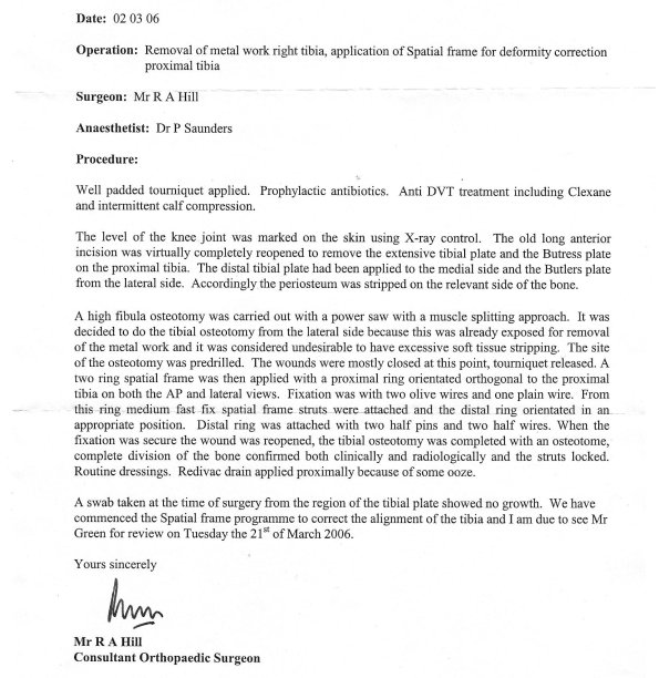
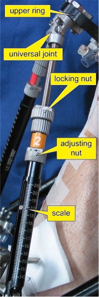
I'm in hospital for 8 nights, and home on 10th March, 2006 (Day 257). The twelve holes, due to the 5 wires which pass through the leg, and the two titanium pins screwed into the leg, need to be cleaned daily. The six TSf struts are adjusted to slowly realign the leg. So, I'm back to the zimmer frame and two crutches...
Adjustment Programme:
Over a period of 65 days, until 12th May, 2006 (Day 320), the six struts are adjusted each night following five different computer generated adjustment programmes.

Click to see adjustment programme #1
Taylor Spatial Frame X-rays and Photos
1st April 2006 (Day 279): Blue Peter Moment
In true Blue Peter fashion, I at last take the risk and start taking a shower by employing a dustbin liner and copious amounts of sticky tape to protect the TSf, and the holes in my leg, from soap and water...
15th May 2006 (Day 323): Continuing Physiotherapy
I start Physiotherapy, with Louise, Deputy Physiotherapy Manager, at The City Sports Clinic, London Bridge Hospital.
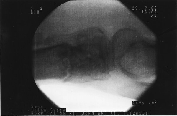
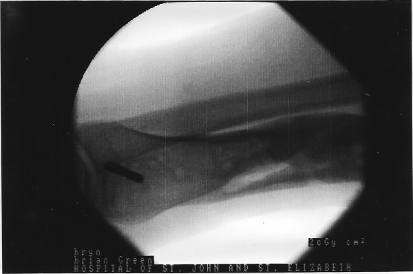
July 2006: Taylor Spatial Frame Removal
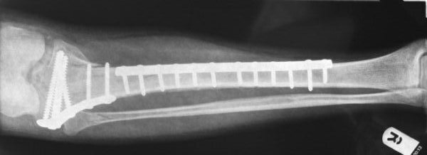
On Saturday 29th July (Day 398) I return to the Hospital of St. John & St Elizabeth (HJE) for a midday 1:15 hour operation on both legs conducted by Mr Hill. Mr Hill removes the Taylor Spatial frame from the right leg and the plate and 12.5 screws (see the x-ray!) from the left leg. I'm home, to start 10 days convalescence, midday on the Sunday.
September 2006: Final Assessment
On 13th September (Day 444) I have a follow up consultation with Mr Hill at The Portland Hospital (for Women and Children). X-rays are taken of both legs - notice the extended muscle on the front of the right leg due to the epimysium being cut during the original operation in France.
So what's the damage?
My right leg has been rotated out to the right by 16°, which is the equivalent to moving the foot 13cm out to the right. It has been rotated forward 10°, from a break 5cm below the knee joint. And, now points an almost normal X° out, after rotating the bone radially through Y°.